

What Is Cataract?
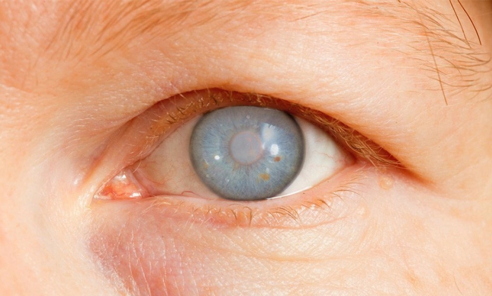
Cataract is opacification or clouding of the crystalline lens . The word “cataract” stems from the Greek word for “waterfall,” because prior to the 1700s, people believed that cataracts were composed of an “opaque material flowing, like a waterfall, into the eye”. It may be congenital or acquired.
How common is Cataract?
Cataract is the commonest cause of visual impairment and blindness worldwide. According to WHO 2010 assessment, cataract is responsible for 51% of world blindness, accounting for 20 million blind people. As people in the world live longer, the number of people with cataract is anticipated to increase (WHO). Cataract was found to be the leading cause of blindness in all the surveys we conducted in various communities. In both Atakunmosa West and Ife North LGAs, cataract accounted for half of the blindness, while in the special education school and among prisoners, it represented a quarter.
Risk factors for developing cataract
Several risk factors have been identified to predispose to cataract formation. These include advancing age, female gender, heredity, steroid drug use, smoking, sunlight exposure and high body mass index (Mukesh et al, 2006).
The prevalence of cataract is higher in diabetics compared to their age-matched counterparts in the same environments. Cataract was the leading cause of blindness (60 %) and visual impairment (59%) in patients with Diabetes Mellitus, according to our study (Onakpoya… .Adeoye et al, 2015).
We conducted a case control study on dietary and lifestyle patterns of 62 subjects made up of 31 patients with cataracts and 31 control subjects without cataracts. The study showed that higher percentages of controls than patients had adequate intakes of fruits and vegetables. Vitamin supplement usage was also higher in the controls than in patients with cataract. We demonstrated a strong association between past history of smoking, alcohol consumption and cataract (Ojofeitimi….. Adeoye et al, 1999).
This brings us to the question, “Can cataract be prevented?” Currently, no study has absolute proof of cataract prevention or slowing of its progression. However, lifestyle changes that take care of the modifiable risk factors may be helpful. These include:
- Stop smoking
- Reduce alcohol intake
- Wear sunglasses
- Control diabetes
- Maintain a healthy weight and
- Eat a healthy diet rich in colorful fruits and vegetables which have many antioxidants to maintain healthy eyes.
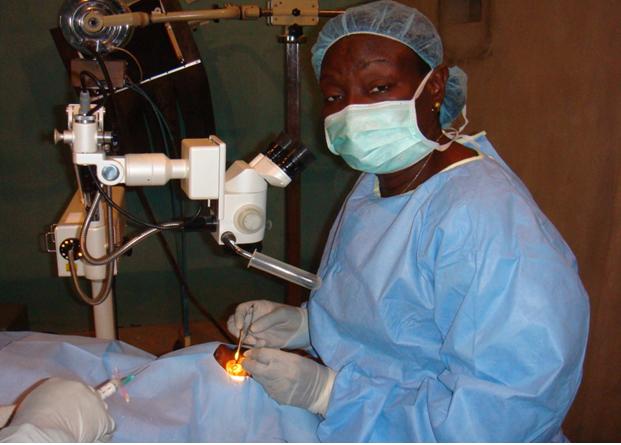
The good thing about cataract is that blindness from this condition is treatable and therefore, reversible. Surgery is the only form of treatment for cataract. Fortunately, this type of surgery is considered to be the most cost effective of all surgical interventions in eye care, in view of the great returns in good vision, if meticulously performed.
Cataract surgery
“The only real voyage of discovery consists not in seeking new landscapes, but in having new eyes…” Marcel Proust.
Before 2000 BC, there was no specific record of eye surgery, but it is possible that Babylonians used digital pressure on the eyeball to dislocate the lens from its zonular attachments .
Early Hindu references to Couching were by Susruta, using sharp needle knives to displace the lens from the pupil into the vitreous cavity (Garg, Das, 2013). This procedure was performed by itinerant surgeons in Europe, Arabia, and Asia. It is still being performed in 21st Century Nigeria by fake eye ‘surgeons’ originally from the North, but now from any part of the country and other West African countries (Osuntokun, 2001). Couching could be successful if the whole cataract is dislodged from the pupil, giving a brighter, but unfocussed image of the object viewed. However, if the lens is ruptured, severe inflammation and infection often lead to blindness. A great breakthrough occurred in the 1940s in England when Harold Ridley invented the intraocular lens (IOL). The IOL is a small, special plastic lens implanted inside the eye to replace the cataract that was removed, making it possible for the operated eye to see clearly after surgery.
In 1995, with the support of Prof. R.O.A. Makanjuola as the Chief Medical Director of OAUTHC, I organized and led the ophthalmic team to conduct the first ever surgical eye camp in rural areas of Imesi-Ile and environ (Fig. 6). The aim was to promote awareness of the community to eye disorders through health education, publicize eye care facilities available in OAUTHC, provide cheap surgical intervention and train community health officers to recognize common eye disorders for early referral. This pilot eye camp metamorphosed into a robust Community outreach programme of the Ophthalmology Department.
In 1999, after attending a skill-acquisition course in intraocular microsurgery at the National Eye Centre, Kaduna, I attracted a collaboration between Sight Savers International and OAUTHC.
This international non-governmental organization donated sets of ophthalmic instruments and consumables for our outreach programmes. Consequent on these outreaches, the Federal Government of Nigeria designated OAUTHC a regional centre for the National Prevention of Blindness Programme.
 What is Glaucoma?
Glaucoma is a group of eye diseases that cause irreversible damage to the optic nerve which carries visual impulses to and from the brain so that one can have good vision. There are many types of glaucoma and oftentimes, the pressure inside the eye rises to a level higher than is healthy for the eye. Glaucoma may result in blindness if not treated. It is the second most common cause of blindness worldwide, especially in many countries in West Africa (Ashaye, Adeoye, 2008). The incidence of glaucoma is estimated to be 5.5/1000 persons/year in Blacks (Dandona, 2001); therefore, in Nigeria, we expect additional 180,000 new cases every year (Ashaye, 2010).
Glaucoma was responsible for 11% of blindness in Ife North LGA, 14% in Atakumosa West LGA, 10% in prisoners and 20% among students of special education school (Adeoye, 1996; Onakpoya, Adeoye…., 2007; Ajite, Adeoye, 2010; Onakpoya….Adeoye, 2011).
The bad thing about glaucoma is that it causes irreversible blindness. The disease creeps in painless and unnoticed until it has blinded at least one eye such that the Yorubas call it “Adake f’oju” (blinds in a quiet manner).
Prevention of glaucoma blindness hinges on early detection, compliance with medication and life-long follow-up. Important challenges in glaucoma management, resulting in progressive loss of vision are non-compliance with medication and drop-out from follow-up. We examined the clinical characteristics of patients who drop out in the first year of follow-up from a glaucoma clinic. The dropout rate was high at 61%. Patients who were more likely to drop out were younger patients, males, those who travelled far distances to the clinic, those with mild to moderate glaucoma, those with no family history of blinding eye diseases, and patients using 2 or more eye drops. Patients who seem to perceive their problems as not serious, dropped out of follow-up. These findings have great implications in planning future studies and intervention to improve the follow-up of glaucoma patients (Ashaye & Adeoye, 2008).
Another reason why patients might stop coming to the hospital is, not observing an improvement in their vision due to irreversible optic nerve damage. Some believe it is a spiritual attack, hence, it needs spiritual management.
A 40-yr-old carpenter who still had some vison in both eyes was diagnosed with glaucoma and appropriate eye drops were prescribed. He attended one follow-up clinic only to default for about 6 months. He returned to the hospital with total blindness in both eyes. On further questioning, he admitted to seeking prayers from one prophet on a mountain top who asked him to lie face up, open his eyes and gaze directly at the sun until sunset for 7 days! Of course, the sun burnt out the fovea, which is the most sensitive part of the eye for seeing (Solar retinopathy).
Indeed, as Martin Luther King Jr. put it,
“Nothing in the world is more dangerous than sincere ignorance…”
Optic atrophy
Other non-glaucomatous causes of optic atrophy leading to blindness that we observed in the communities include trauma, intracranial tumours, optic neuritis, Leber’s hereditary optic neuropathy and compressive optic atrophy in patients with Burkitt’s lymphoma (Adeoye et al, 2007; Adegbehingbe…Adeoye, A.O et al, 2005).
- Gender issues
The bad aspect relating to gender is that two-thirds of blindness and visual impairment occur in women (Clayton & Davis, 2015). Even in this 21st century, women still have to depend on their husbands for decision-making regarding their eye health care. In a recent study we conducted, we found that males that fully self-funded their eye care services were significantly more than females (52% vs 28%) while monthly average personal income was not significantly different between the genders. The study also showed that more females consider their spouses the most influential individual involved in the process of taking health-related decisions (Adeoye, Awe, 2016).
What are the causes? It is known that cultural patterns and social factors within the household and community favour males. Also, women have a low level of control over household resources. Poverty and visual impairment have long been reported to be closely interrelated such that either acts as a cause and effect of the other (Jaggernath et al, 2014). In Nigeria, a significant proportion of the population lives below the poverty threshold, thereby limiting their access to healthcare and other basic needs. Also, women’s child care responsibilities may make it difficult for them to leave home. These issues underscore the importance of outreach programmes to bring eye care closer to the people.
ORBITO-OCULAR DEFORMITIES AND MASSES
The face, including eyes, is first viewed as a person approaches. Abnormal growths and deformities of the eye and orbit may be congenital (i.e. developmental defect before birth) or acquired (disorders that develop after birth). They can make an individual look ugly and provoke a negative attitude from members of the community, as Yorubas believe in the “normal” and “perfect” and abhor the “abnormal” and “imperfect”, avoiding sustained social interaction with them. (Togonu-Bikersteth & Odebiyi, 1985).
Cases seen and reported include, isolated bilateral congenital coloboma (defects) of the upper lid, associated with symblepharon in a 5-month old baby (Adegbehingbe, Olabanji, Adeoye, 2005) and congenital bilateral sporadic aniridia (absent/ rudimentary iris) in a 5-year old girl (Adeoti, Ashaye, Adeoye, 2010).
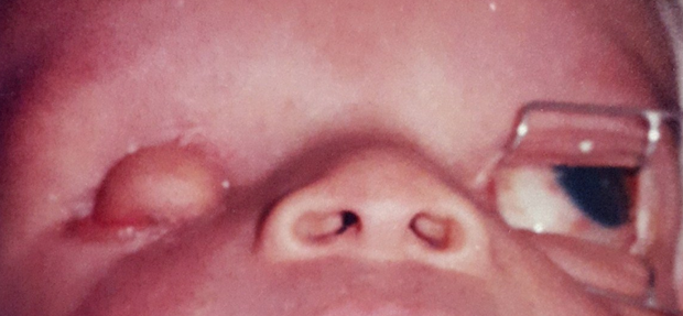
A case of Goldenhar’s syndrome, otherwise known as oculoauriculo-vertebral dysplasia was documented in a 3-day old neonate. This is a rare congenital complex of abnormalities involving the eyes, ears, spine, and jaw. The mother had ingested some traditional medicine concoction when she was 3months pregnant as a prophylaxis against recurrent abortion. The baby was born with right anophthalmos (absence of eyeball). The empty orbital space was filled with a soft, solid mass with skin protruding from the socket and attached to the tarsal conjunctiva of the lower lid. The left eyeball was normal but had a limbal dermoid cyst on the temporal aspect. He had abnormal skin tags in front of both ears and small, maldeveloped lower jaw. The aetiological factor was presumably the traditional medication as it has been reported that maternal drug ingestion within the first trimester of pregnancy may predispose to Goldenhar’s syndrome. The importance of health education to pregnant women in order to prevent congenital abnormalities in the newborn can not be overemphasized (Adeoye, 2002).
Eye injuries
Despite the protected position of the eyes within the bony orbits, they are still vulnerable to injury because of their anterior exposure. Eye injury is by far the commonest cause of blindness and visual impairment in at least one eye. It is the leading cause of eye removal (Adeoye & Onakpoya, 2007; Awe, Adeoye et al, 2016). There is a general assumption that eye injuries are accidental and out of human control; but, they are preventable by taking extra care. Globally, it is estimated that injuries were responsible for 1.6 million people blind in both eyes and 19 million people blind in one eye (Negrel, 1998). Our studies revealed that eye injuries were responsible for 55% of all emergency eye consultations in OAUTHC (Onakpoya & Adeoye, 2008).
Eye injuries in the young
The studies we conducted on children and young adults showed that 1 in every 5 children presented to the Eye clinic with eye injury, which was more common in males (Adeoye, 2002). Injuries occurred with increasing frequency after the age of 4 years and children were commonly injured at play (50%), during corporal punishment (10%), assault (9%), and street hawking (9%). The causative agents were mostly sticks and twigs, followed by missiles and falls (Adeoye, 2002; Onakpoya & Adeoye, 2009).
A common form of missile in schools these days is the catapaulted broomstick with sharpened point, slung and propelled with a rubber band, which usually causes penetrating eye injury with retained bits of broomstick in the eye. The broomstick being an organic matter, provokes a fungal eye infection that may completely destroy the eye.
Visual prognosis was best in patients with closed-globe injuries (48%) and worst in those with open-globe injuries (31%).
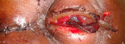
Severe open-globe injuries with globe rupture often necessitate eye removal. Figure 9 shows the eye of a 40-year old man who attempted to inspect his car engine while the engine was running, during repairs. The fan blade came off its hinge and hit his left eye and nasal bridge, resulting in a completely disorganized eyeball with loss of eye contents. This necessitated an emergency eye removal.
Eye injury in times of peace and conflict
In times of peace, the economic downturn of many families has necessitated game hunting by untrained hands, using crude locally manufactured Dane guns.
I observed from my study, that severe open-globe injuries associated with retained intraocular/orbital foreign bodies resulted from such guns with unstable parts. Loose metallic burner, would come off and hit the aiming eye, often necessitating eye ball removal (Adeoye, 1996).

The eyes have greater risk of injury in conflict than other body parts due to the preferential exposure of the face in combat. We conducted a study of all patients with eye injuries resulting from the Ife/Modakeke communal conflict, treated at Obafemi Awolowo University Teaching, Hospital, Ile-Ife and 2 private eye clinics to determine the cause, morbidity and visual outcome of ocular injuries sustained. Fifty-five injured eyes of 54 patients were studied. The mean age was 32 years with a male preponderance of 96%. Forty-two eyes (76%) were injured by gunfire through direct impact, backfire or stray bullet (Fig 13). Thirty-one eyes (56%) sustained closed-globe injuries while 24 (44%) had open-globe injuries. Thirty-five eyes (64%) were blind, out of which 9 eyes (16%) required primary eye removal (Adeoye et al, 2002).
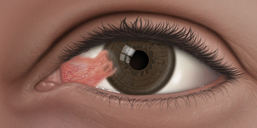 PTERYGIUM AND PINGUECULUM
Pterygium
Pterygium (pronounced tur-IJ-ee-um) is a common eye condition that affects people who spend a lot of time outdoors. People with pterygium have a growth of pink, fleshy tissue on the white of the eye. It usually forms on the side closest to the nose.
It is a noncancerous lesion that usually grows slowly throughout life. Or it may stop growing after a certain point. In rare cases, a pterygium can continue growing until it covers the pupil of the eye and interferes with vision.
A pterygium may affect one or both eyes. When it affects both eyes, it is called a bilateral pterygium.
Pterygium is usually not a serious condition. But it can cause annoying symptoms such as a feeling of a foreign body in the eye
Causes
- Prolonged exposure to ultraviolet light
- Dry eye
- Irritants such as dust and wind
It’s most often seen in young adults’ ages 20 to 40. It appears to be more common in men than in women.

Right pterygium
Symptoms
- Burning
- Gritty feeling
- Itching
- Sensation of a foreign body in the eye
- Blurred vision
Treatment
See an ophthalmologist if you have symptoms of pterygium. Pterygium usually doesn’t require treatment if symptoms are mild. If a temporary worsening of the inflamed condition causes redness or irritation, it can be treated with:
- Lubricating eyedrops or ointments
- Occasional use of vasoconstrictor eyedrops
- Short course of steroid eyedrops
If the lesion causes persistent discomfort or interferes with vision, it can be surgically removed during an outpatient procedure. You and your doctor may also take into account appearance and the size of the pterygium when making a decision about surgery.
Pinguecula

A pingueculum is a small, yellowish bump on the conjunctiva near the cornea. It can appear on either side of the cornea, but occurs more often on the nose (nasal) side. The growth may increase in size over many years.
Pingueculae can occur at any age, but they’re mainly found in middle-aged and elderly people.
Causes
It’s been linked to frequent exposure to sunlight, dust, or wind
Symptoms
Pinguecula will rarely cause symptoms but they can cause irritation if they become elevated.
- Being yellowish in color
- Itching and irritation
- Having dry eyes
- Redness when irritated
- Gritty feeling or foreign body sensation.
Treatment
Because they are benign tumors they will usually not require any type of treatment. If Pinguecula becomes inflamed and causes dryness your ophthalmologist or physician may prescribe artificial tears to help lubricate your eyes.. If the eye appears to be swollen you may be prescribed a mild anti-inflammatory medicine to help reduce it.
The ophthalmolgist may prescribe mild steroid eye drops to be used on a temporary basis.
It is very rare that a person has to have surgery but if the appearance of the Pinguecula bothers you, the ophthalmologist could remove it.
 It involves inflammation of the outer layer of the eye and inside of the eyelid. Eye redness may also be due to swollen or dilated blood vessels, which cause the surface of the eye to look red, or bloodshot. It is often times an ophthalmologic emergency.
Causes include:
- Bacterial or viral infection- can be spread from person to person
- Allergies
- Blepharitis — Swelling of the eyelash along the edge of the eyelid.
- Conjuctivitis — Swelling or infection of the tissue that lines the eyelids and coats the surface of the eye (the conjunctiva). This is often referred to as “pink eye.”
- Corneal ulcers– Ulcers on the outer covering of the eye, usually because of a bacterial or viral infection.
- Uveitis — Swelling of the uvea, which includes the iris, ciliary body, and choroid. This is often related to an autoimmune disorder, infection, or exposure to toxins. Often, only the iris is inflamed, which is called iritis.
- Contact lens products, eye drops, or eye ointments
- Straining or coughing This can lead to a bright red, dense bloody area on the white part of the eye. This is called a subconjunctival hemorrhage
It can cause swelling, itching, burning, discharge, sensitivity to light and redness
TREATMENT
It is important to visit an eye doctor so as to establish a diagnosis because treatment depends on cause.